is left ventricular hypertrophy life threatening
HOCM is an autosomal dominant genetic disorder in. Annualized continuous flow left ventricular assist device CF LVAD implants from 2011 to 2020.

Hypertrophic Cardiomyopathy In Children Cs Mott Children S Hospital Michigan Medicine
Ventricular arrhythmias can occur without warning at almost any level of hyperkalemia with normal EKG.

. Atrial Fibrillation EP-IA Clinic. The ventricular rate is typically very high 100250 beats per minute and cardiac output is affected ie reduced in virtually all cases. The patient had malignant.
Ventricular fibrillation is a chaotic rhythm where the ventricles quiver rapidly in a purposeless fashion. When LVAD patients are stratified by number of readmissions during the first 6 months after implant we see significant differences in survival. Far-field activation was equally early 30 ms ahead of surface QRS in regions of the left coronary cusp-right coronary cusp LCC-RCC right ventricular outflow tract RVOT and epicardially in great cardiac vein-anterior interventricular vein GCV-AIV.
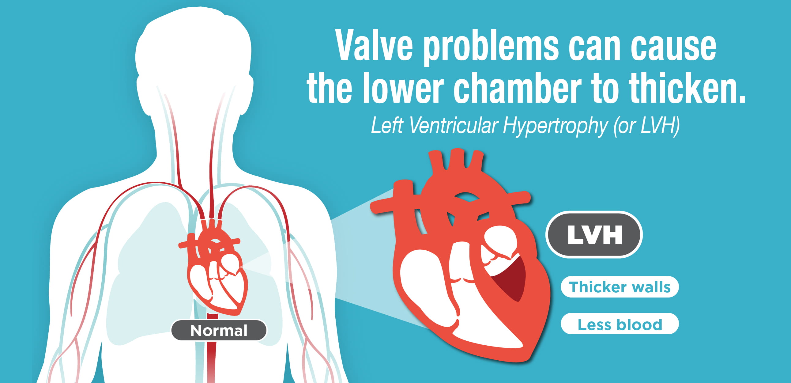
If LVH enlarges your heart it can compress the. In 2020 there was 17 reduction in volumes with 83 of patients receiving a fully magnetically levitated centrifugal-flow device. Ventricular tachycardia cause immense strain on the ventricular myocardium simultaneously.
Ventricular tachycardia is life-threatening because. While it is true that right ventricular hypertrophy can be life threatening there are literally thousands of people with the condition that are. Incidence of Myopericarditis and Myocardial Injury in.
This change is reflected in the appearance of the QRS complex of the ECG. At serum potassium level 80 mEqL the P wave disappears the T waves are fused with the QRS complexes culminating in a sine-wave rhythm. HOCM can lead to clinical heart failure life-threatening arrhythmias mitral regurgitation and sudden cardiac death.
And left ventricular hypertrophy LVH predispose to ventricular arrhythmias polymorphic ventricular tachycardia VT and VF. Atrial fibrillation affects the upper chambers of the heart known as the atria. Atrial Ectopic Tachycardia EP Clinic.
Premature ventricular complexes PVCs originating from region of left ventricular LV summit. VA include a spectrum that ranges from premature ventricular complex PVC to ventricular fibrillation VF with a clinical presentation that ranges from a total lack of symptoms to cardiac arrest. The relative right ventricular hypertrophy of the neonate regresses over the first few months of life.
LVH creates a more static EKG pattern over time whereas Wellens A should always evolve. When you have CHD your heart must work harder. It frequently develops into ventricular fibrillation.
How to identify patients at high risk of SCD with HF and LVH is uncertain. When it detects a very fast abnormal heart rhythm it delivers energy a small but powerful shock to. 50 Changes in left ventricular geometry affect the likelihood of developing VT and VF.
Apparent Life-Threatening Event UCP Clinic Arrhythmia Anything except sinus rhythm Possibly EP Clinic. International Consensus on Cardiopulmonary Resuscitation and Emergency Cardiovascular Care Science With Treatment Recommendations. Atrial Flutter EP Clinic.
This is a life-threatening situation because can degenerate into ventricular fibrillation or asystole. Full length article. Guideline Directed Medical Management of Heart Failure with Reduced Ejection Fraction.
Most life-threatening VA are associated with ischemic heart disease particularly in. The heart with ventricular fibrillation cannot pump blood effectively to the brain and the rest of the body. ICDs are suggested for people at risk for life-threatening arrhythmias or sudden cardiac death.
The ICD is a small device placed just under the skin and is connected to wire leads that are threaded through the vein to the heart. There is mild asymmetric hypertrophy basal septum 14 cm. Fibrillation can affect the atrium atrial fibrillation or the ventricle ventricular fibrillation.
Regardless of etiology and ECG ventricular tachycardia is always a potentially life-threatening arrhythmia which requires immediate attention. Guidelines summarize and evaluate all available evidence on a particular issue at the time of the writing process with the aim of assisting health professional. This electrocardiogram ECG shows evidence of severe left ventricular hypertrophy LVH with prominent precordial voltage left atrial abnormality lateral ST-T abnormalities and a somewhat leftward QRS axis 15º.
Systolic function is hyperdynamic with an ejection fractio. Patients may be asymptomatic or may present with life-threatening hemoptysis resulting from bronchial artery hypertrophy dilatation and eventual rupture. Long-term outcomes in inferior ST-segment elevation myocardial infarction patients with right ventricular myocardial infarction.
View all 2020 CoSTR Papers. The characteristic CT appearance of mycetoma is a solid round or oval gravity-dependent mass within a. Resuscitation is a monthly international and interdisciplinary medical journal.
Right Ventricular Dysfunction in Critically Ill Patients With COVID-19. ECG typical findings in resting position are for example sinus bradycardia atrioventricular block primary and secondary and right. The most significant of all rhythms associated with heart failure are the life-threatening ventricular arrhythmias.
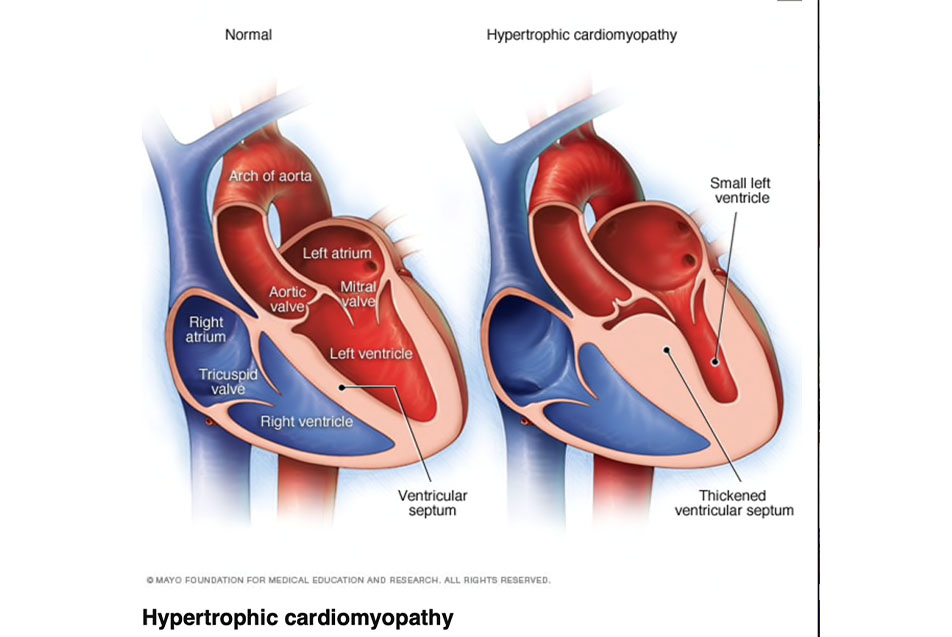
Left ventricular hypertrophy regression No left ventricular hypertrophy regression The medical history of the patient endurance sports and physical examination bradycardia and maybe a third or fourth heart sound can give important hints. Life-threatening features in an unstable patient include. Asymmetric septal hypertrophy is the most common type of hypertrophic cardiomyopathy in which the abnormal ventricular muscle thickening is confined to the interventricular septum causing the walls of the lower heart chambers typically the left ventricle to become thick and stiff 1The hypertrophy in this phenotype of hypertrophic.
Ablation from all these sites in this. LVH tends to cause TWI in a more lateral distribution V3-V6 than Wellens V2-V4. Often occurs in the context of benign early.
Left ventricular hypertrophy LVH LVH can very closely mimic Wellens creating a pseudo-Wellens pattern. CHD can cause LVH and vice versa. Ventricular fibrillation is imminently life-threatening.
Atrial fibrillation may be due to serious underlying medical conditions and should be evaluated by a physician. Atrial Septal Defect. Right Ventricular Failure Manifesting in COVID-19 ARDS.
The mean frontal plane QRS axis of the neonate is around 75 with a range from 60160. The left ventricular cavity appears normal size. An ICD constantly monitors the heart rhythm.
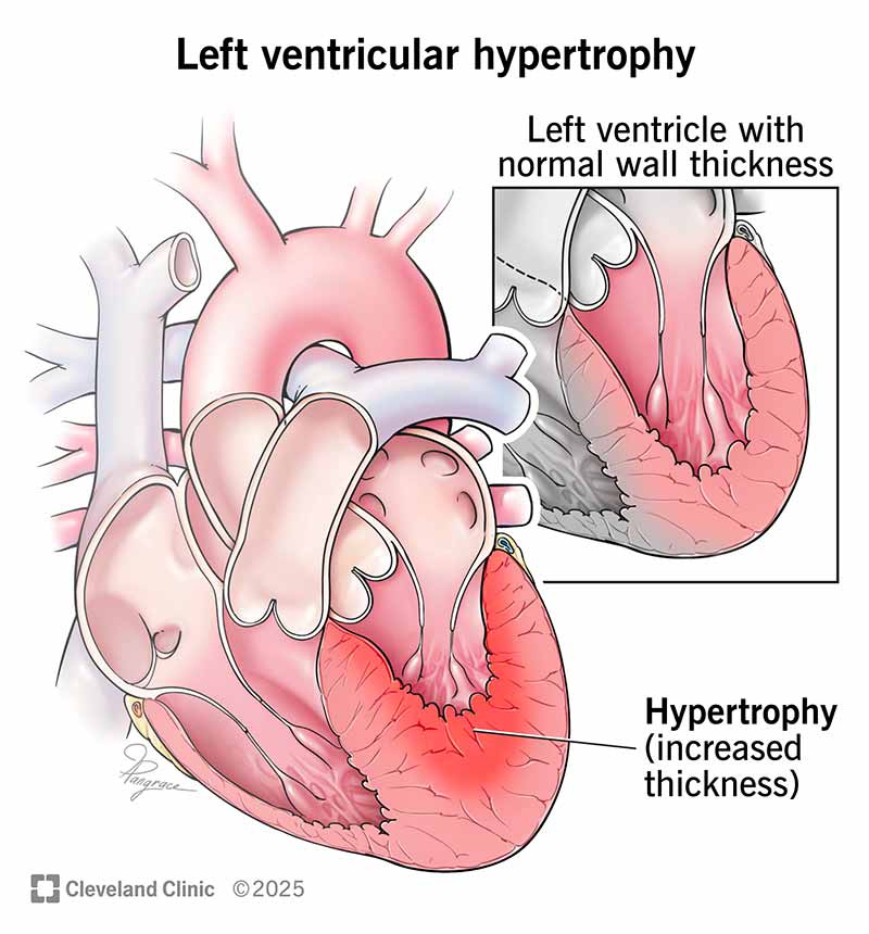
The condition is known as left ventricular hypertrophy LVH. A Call to Transition from VV-ECMO to RVAD-ECMO. There is a relatively rapid change in axis over the first year of life and from this age onwards the mean frontal QRS axis.
Left atrial strain in cardiac surveillance of bone marrow transplant patients with prior anthracycline exposure. Long-Term Impact of Preventive Tricuspid Valve Annuloplasty on Right Ventricular Remodeling. Adult Basic Life Support.
My echocardiogram results says. It occurs suddenly with no prior warning and. ARVD AKA ARVC Arrhythmogenic Right Ventricular Dysplasia UCP Clinic.
Improved Outcomes with Quadruple Therapy.

How Is Left Ventricular Hypertrophy Treated

Left Ventricular Hypertrophy Enlarged Heart Vascular Health Clinics
What Does Mild Conc Lvh Means Is It A Big Disease Does It Cause Heart Attacks Quora
What Is Left Ventricular Hypertrophy Lvh American Heart Association
Hypertrophic Cardiomyopathy Hcm Symptoms Causes Heart Foundation

Left Ventricular Hypertrophy Lvh Symptoms Causes And Treatment
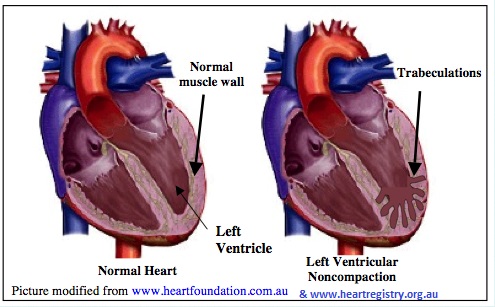
Left Ventricular Non Compaction Cardiomyopathy Cidg
Left Ventricular Hypertrophy Lvh Causes Symptoms And Treatment
Your Symptoms Could Be A Sign Of Hypertrophic Cardiomyopathy